The first time my medial collateral ligament was injured
About 20 years ago I had a motorcycle accident that resulted in the bike landing on the inside of my knee. I never had it x-rayed but by the location of my subsequent knee pain that ligament seemed to the most likely victim. For quite a while afterwards, at various times, I could be walking and my shin felt like it came loose from my femur, like the tendons and ligaments hand jumped out of their tracks. I'd then have to try to wiggle or jiggle things back into place.
The loose knee took time to heal. I never really figured out any special techniques for holding it in place. But even after that sort of symptom disappeared, there where certain things that bothered that knee, even until recently.
I then experience similiar symptoms with the other knee. One of the problems this time was that I was teaching yoga classes and when I teach I demonstrate most of the poses. I found some muscle activation techniques that prevented knee pain. After several years of study I think I understand the mechanism that helped prevent medial collateral knee pain.
Attachments of the Medial Collateral Ligament
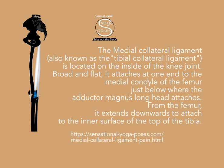
The Medial collateral ligament (also referred to as the "tibial collateral ligament") is located on the inside of the knee joint.
Broad and flat, it attaches at one end to the medial condyle of the femur just below where the adductor magnus long head attaches to the adductor tubercle.
From the femur, it extends downwards to attach to the inner surface of the top of the tibia.
At the tibia, the posterior fibers angle backwards and insert just above a groove where the semimembranosus muscle attaches.
It passes over the front front part of the semimembranosus tendon and has a few fibers that attach to this tendon.
The bursa semimembranosus is sandwiched between the medial collateral ligament and the tendon of the semimembranosus muscle.
The anterior fibers of the medial collateral ligament angle forwards and attach to the top of the shaft of the tibia, just below the bulge of media condyle. Here it is positioned underneath the pes anserinus, which is the joined tendons of the sartorius, gracilis and semitendinosus muscles.
The anserine bursa lies between the medial collateral ligament and the tendons of the sartorius, gracilis and semitendinosus.
The medial collateral ligament represents the tail end of the adductor magnus muscle (the long head.)
Some "Obvious" Muscle Activations for Dealing with Medial Collateral Ligament Pain
Because of the close relationship with the adductor magnus long head, semimembranosus, semitendinonosus, gracilis and sartorius, one possible starting point for dealing with medial collateral ligament pain is to activate these muscles.
When playing with "muscle activation" or more generally, "muscle control", it's important to realize that muscles have to work against an opposing force to activate.
If you activate a muscle while restricting movement then the muscle you are activating is working against an opposing muscle so that it can activate without causing movement. As an example of this, if you tense your biceps, your triceps will also activate.
The result is that not only do you make your biceps "pop", you also stabilize the elbow joint.
Adductor Magnus Long Head Activation
The adductor magnus long head can be used to create an internal rotation of the femur relative to the pelvis. To activate it while standing, try pressing back on the inner thigh while preventing the femur from rotating. Also prevent your pelvis (and lower leg) from moving.
If you successfully activate the adductor magnus long head, then external rotator muscles will also activate to stabilize the femur against rotation. You'll probably feel your gluteus maximus activating to resist the internal rotation causes by adductor magnus long head.
With adductor magnus activated you can note for yourself whether this decreases medial collateral ligament pain. You can try to maintain the activation and vary the rotation of your thigh to see if that helps. This can be likened to turning a guitar.
If you get medial collateral ligament pain while walking, you could try to activate your adductor magnus as your front foot touches the ground.
So that you can comfortably maintain adductor magnus activation you may have to stabilize your heel against side to side rolling. (Read Shin Rotations and Heel Stability for more on how to stabilize the heel against side to side rolling.)
Pes Anserinus Sartorius Gracilis and Semitendinosus
The pes anserinus muscles run up the inner thigh from the tibia to attach to prominent points of the pelvis:
- The Sartorius to the ASICs,
- the gracilis near the pubic bone and
- the semitendinous to the Ishcial Tuberosity or sitting bone.
These muscles can be used to rotate the shin inwards when the knee is bent. If the knee is straight the sartorius can be used to rotate the shin (and femur) outwards.
With the knee stabilized, the sartorius can aslo be used to flex the hip (bent the hip forwards.)
More "Long" Hip Muscles
The iliotibial band runs up the outside of the thigh and attaches to the top of the outer edge of the tibia, just in front of the fibula. The tensor fascia latae attaches to the front edge of the IT band from the ASICs while the gluteus maximus attaches to the back edge.
These muscles can also be used to rotate the shin inwards and outwards.
The outer hamstrings muscles (the biceps femoris) attach to the top of the fibula. These too can drive external rotation of the shin when the knee is bent.
Used against each other these "long hip muscles" can be used to stabilize the shin against rotation.
Reducing Slack with the Vastus Muscles
Apart from creating stability at one end of a muscle, another important aspect of muscle control is reducing slack (or creating it) so that a muscle can activate effectively.
To that end, the vastus muscles can be used to take out slack from some of the long hip muscles. For example, the vastus lateralis can activate to take out slack from the IT band. The vastus intermedius can take out slack from the rectus femoris which in turn may also take out slack from the sartorius. Sartorius and gracilis may also be tightened slightly by activating the adductors as well as the vastus medialis.
And so an important ingredient for long hip muscle activation is taking out their slack via activation of the bulkier thigh muscles over which they run.
Stabilizing the Shin via Lower Leg Muscles
Most of the methods described so far are muscle control methods for invoking stability.
The first method, using the adductor magnus, stabilize the thigh relative to the pelvis. The second method stabilizes the shin relative to the pelvis.
Another possibility is to stabilize the shin (and heel) against rotation via the foot and ankle.
With your foot and lower leg stabilized you could then experiment with activating the pes anserinus muscles (together or individually) to see if that alleviates medial collateral ligament pain.
The Gastrocnemius
The gastrocnemius is the large fo the two calf muscles. It's tendons interlace with the hamstring tendons, including the semimembranosus and semitendinosus. Activating the calf may add tension to the tendons of the semitendinosus and semimembranosus.
It's a simple way to stablize the knee and you may find that on occasion it helps you to deal with medial collateral ligament pain.
Stabilizing the Hip Bone
Since the aforementioned long muscles of the hip attach to the hip bone, another strategy is to try to stabilize the hip bone. Three long hip muscles attach to the ASICs or there abouts, the sartorius, tensor fascia latae (and IT Band) and the rectus femoris. To give all three of these muscles a stable foundation try creating an upwards pull on the ASIC. You may find it helpful to lift your ribs, in particular your lower back ribs. You may then find it easier to use the external obliques to create an upwards pull on your ASICs. Note that the goal isn't to actually cause the ASICs to move upwards, just create an upwards pull on it so that it resists being pulled downwards. On occasion you may find this sufficient. However, you may find that on some occasions it helps to also activate the vastus muscles so that not only are you giving these long hip muscles a stable foundation, you are also taking out the slack.
My Story
When I had a motorcycle accident about 18 years ago I'm pretty sure it damaged my Medial collateral ligament. It hurt to put pressure on it for a while. Then it got a little bit better but then the problem would be that my knee felt like it cam out of alignment, like it was loose and I somehow hand to shake or adjust it back into alignment. It was very frustrating.
Fifteen years later I damaged my other knee. The pain was almost the same. And the knee "falling out of joint" was similiar too. I live in Taiwan and I went to see the local equivalent of a chiropractor a couple of times to get the knee treated, which was helpful on a couple of occasions but I think that a big part of my knees, both of them, getting back to full operability was time, but also muscle control. Playing with different muscle activations to see what works. One of the benefits, apart from making my medical collateral ligament pain go away is that some of the same muscle control techniques I developed for dealing with pain also helped me to improve my flexibility.
I've also gotten to better understand my body (and bodies in general) in the process. I'd suggest that if you want to fix your knee yourself then learn how to control your muscles. Muscle control is not only activating muscle but relaxing it. It's also the mechanism by which you feel your body. If you haven't got muscle control then you can't feel your body. For an introduction to muscle control that focuses on the legs, check out sensational leg anatomy. You'll learn most of the activations talked about in this article.
Keys to Understanding Medial Collateral Ligament Pain
With respect to medial collateral ligament pain, (pain at the inside of the knee) it may be helpful to understand the movement possibilities of the tibia and fibular relative to the thigh and hip bone and to also understand how the sartorius, gracilis and semitendinosus can be used to stabilize the shin or, if the shin is stabilized, to stabilize and control the pelvis.
Another important idea is that ligaments are active structures that are affected by muscle tension.
Further to this is the idea that joint capsules are designed to maintain space between the bones that they connect by pressurizing synovial fluid. It's for this reason that the bursae of the knee may be relevant.
Since muscles can affect ligament tension, one other point to note is that for muscles to activate effectively, they may need to be lengthened. You could think of this as "taking out the slack." The vastus muscles, as well as the "wide" portion of the adductor magnus may be important in this regard.
For joint integrity, joint capsules rely on muscle control to vary ligament as well as tendon tension. This tension can be used to directly control joint capsule tension and thus fluid pressure.
If some muscle or set of muscles is not working correctly, the the brain utilizes other muscles to maintain integrity of the joint.
Over use of these "substitute muscles" may result in pain.
Basic Knee mechanics
When the knee is bent the shin can rotate relative to the knee.
Standing with knees slightly bent, the long muscles of the feet can be used to rotate the shins at the same time causing the inner arch of the feet to lift (during external rotation) or lower (during internal rotation.)
Shin rotation can also be caused by the sartorius, gracilis, semitendinosus (which are situation along the inner thigh) along with the tensor fasciae latae and gluteus maximus via the Iliotibial band (which is situated along the outer thigh).
With the knees straight, the shins can't rotate relative to the femurs. The two become locked together.
- Here again the long muscles of the feet and lower legs (tibialis anterior and posterior, peroneals, extensors and flexors of the toes) can be used to rotate shin and femur together.
- The single joint hip rotator muscles could also be used.
- Or the afore mentioned long hip muscles (sartorius, gracilis, semitendinosus, tensor fasciae latae, gluteus maximus via the IT band) can be used.
If there is pain the medial collateral ligament, since it lies close to or connects with the pes anserinus muscles as well as the semimembranosus and adductor magnus, some muscle activation candidates for relieving pain include these muscles.
Sartorius and Gracilis Activation
So that sartorius and/or gracilis can activate effectively, you may find it helpful to activate the vastus medialis muscle and/or the adductors.
Anchoring the Sartorius
An alternative for effective activation of the sartorius is to create an upwards pull on the ASICs. You could think of this as creating an anchor or foundation for the sartorius.
One alternative is to pull upwards by using the external obliques. Try lifting your ribs, in particular your lower back ribs (11 and 12) prior to pulling up on your ASICs.
Another way to create an upwards pull on the ASICs is to create a downwards pull on the same-side sitting bone.
Note:
Same side = Ipsilateral while opposite side = Contralateral.
Using the Semimembranosus
If none of the previous suggestions worked, try activating the semi-membranosus muscle.
One way to add tension to the semimembranosus muscle is to engage the inner/medial head of the gastrocnemius muscle.
Foot and Heel Stability
You may find that in some cases, stabiling your foot, even stabilizing your heel, can help alleviate medial collateral ligament pain. For more on foot and lower leg stability read The foot
For instruction on how to activate muscles of the legs check out sensational leg anatomy.
Published: 2018 06 13


