The Accromion Process
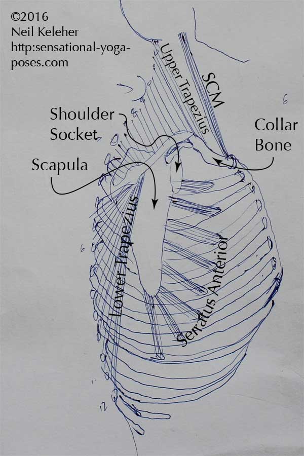
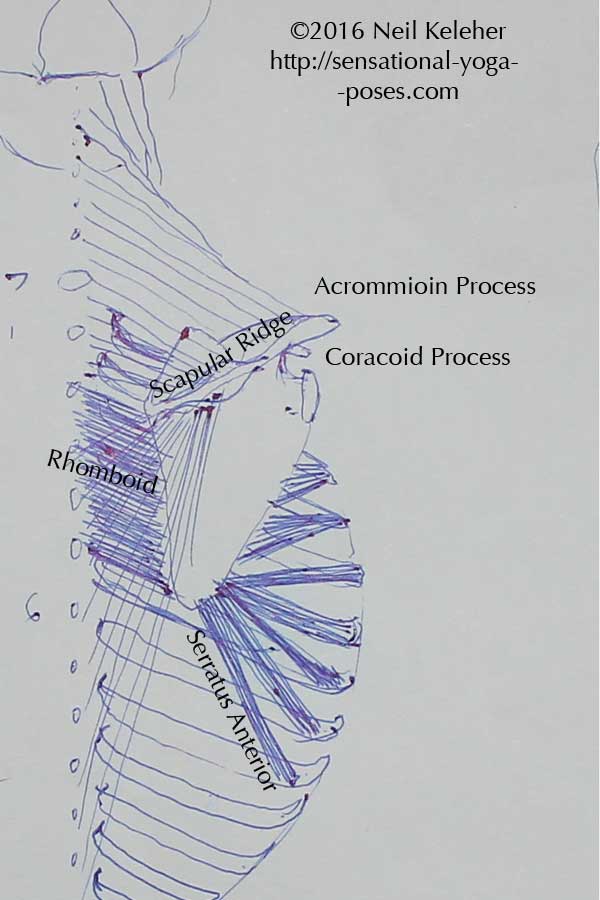
If you feel the top of your shoulder you can feel a peak or crown of bone there. This is the accromion process that marks the end of the scapularridge. For myself I can feel the edge of this projection past the outside edge of my upper arm.
This is where the collar bone attaches to the shoulderblade so it's a good landmark for feeling the connection of the shoulder blade and collar bone. Since the collar bone has the Latin name clavicle, the place where they meet can be called the scapular clavicular joint.
(Note that while muscles act on both the clavicle and the sternum there are no muscles that act across the clavicular joint. While it is helpful to understand that there is mobility between these two bones, it may be helpful to think of the clavicle as an extension of the shoulder blade. It connects it to the sternum.)
Upper Fibers of Trapezius
Attaching to both the accromion process and the outer 1/3 of the collar bone are the upper fibers of the trapezius. These fibers pull upwards on the shoulder blade quite close to the shoulder socket.
You can deliberately activate these fibers by pulling up on the outer end of the shoulder blades (at the accronion process) and the outer end of the collar bones.
The Scapular Ridge
One of the most obvious landmarks of the scapula is the "spine" or "scapular ridge." This is the ridge of bone of bone that runs horizontally across each shoulder blade. The accomion process could be thought of as the extension of this ridge over the shoulder joint.
Towards the inner edge of the shoulder blade its height reduces so that it blends smoothly with the inner edge of the shoulder blade.
Trapezius Lower and Middle Fibers
The trapezius tubercle is a thickening of the scapular ridge close to the inner edge of the shoulder blade. The lower fibers of the trapezius attach to this point. This is the portion of the trapezius muscle that can pull downwards and inwards on the inner portion of the shoulderblade.
Moving further outwards along the top of the ridge of the shoulderblade, the middle fibers of the trapezius attach to the portion of the ridge between the tubercle and the accromion process.
For more on the Trapezius Muscle read:
Trapezius Muscle
The Deltoid Muscle
Another muscle that attaches to the spine of the shoulderblade and the collar bone is the deltoid. Like the trapezius it can be divided into three parts and like the latter muscle it too attaches to the spine of the scapula as well as the collar bone.
The posterior (rear) head of the deltoid could be thought of as a continuation of the middle fibers of the trapezius, reaching down from the outer portion of the scapular ridge to attach to the arm bone.
The Medial (middle) and Anterior (front) heads of the deltoid can be thought of as continuing from the upper fibers of the trapezius. These fibers combine with those of the rear head to attach to the deltoid tuberosity, a bump on the outer edge of the upper arm bone. This "bump" is at about the same height as the bottom of the shoulderblade.
Coracoid Process
Another important landmark on the shoulderblade is the coracoid process (or crows beak.) It's like a finger of bone that sticks out from the top of the shoulderblade reaching just over the front of the shoulder socket.
If you palpitate the peak of the shoulder and then move downwards and then inwards, feeling under the collar bone you may be able to feel a "nub" of bone sticking out about an inch and a half or two inches inwards from the outside edge of the accromion process. That just might be the coracoid process.
Pectoralis Minor, Coracobrachialis, Biceps Brachialis
The coracoid process has three muscular attachments. The pectoralis minor attaches from here to the third, fourth and fifth ribs. This helps to pull the top of the scapula forwards and down. I think of it as helping to flip the shoulderblade over the ribcage.
For more on the Pectoralis Minor read:
Pectoralis Minor
The coracobrachialis attaches from the coracoid process to the upper arm bone, one of the single joint muscles of the shoulder joint.
The long head of the biceps brachialis also attaches to the coracoid process. The short head of the biceps attaches to a tubercle above the shoulder socket or glenoid fossa. The other end of the muscle attaches to the radius and to the fascae covering the flexors of the wrist and fingers.
Attaching the Long Head of the Triceps
Note that a corresponding point on the shoulderblade could be a point downwards and inwards from the shoulder socket.
This is where the triceps long head attaches. From here it reaches down to the point of the elbow, the olecranon process (point of the elbow) of the ulna.
The Inner Border of the Shoulderblade
One of the parts of the shoulderblade that you may be least aware of but that is most useful for learning to control both the serratus anterior and the rhomboids is the inner border of the shoulder blade.
To help students become aware of the shoulder blade in general I have them moving their shoulders back and forwards slowly, so that they can feel their shoulders. For the rhomboids I have them focus on retracting the shoulder blades. For the serratus anterior I have them focus on protracting or spreading them.
In each case I have them direct their awareness to feeling the inner edges of their shoulder blades as they move their shoulders.
Serratus Anterior
The serratus attaches to the front side of the scapula along the inner edge, while the rhomboids attach along the back side of the scapula along the inner edge.
The front side of the inner edge can be divided into three regions, an upper and lower region, both of which are like elongated triangles. Then there is the middle region between them.
For more on the Serratus Anterior,
Including Exercises for Activating it read
Serratus Anterior Muscle
Rhomboid Major and Minor
If you've read anatomy trains then you may have read about how the rhomboids and the serratus are like one continuous muscle with the edge of the scapula glued somewhere in the middle. Where the serratus fibers angle outwards and downwards from the inner border of the shoulder blades, the rhomboids angle inwards and upwards from there towards the spine.
They act to pull the shoulder blades inwards, towards the spine, and upwards also. I'd suggest that these muscles can be useful when pulling the arms backwards behind the body from a downwards position. So if arms are down by your sides, use these muscles to pull your shoulder blades together so that it is then easier to move your arms straight back and up.
Note that when lifting the arms higher behind your back, (such as in prasaritta padottanasana c) you may find it helpful to use the pectoralis minor muscles to pull down on the coracoid process so that the top of the shoulder blade moves forwards.
Another point to note is that if you carry a single sided shoulder bag, you may notice that the pec minor on that side may tend to be overactive. (One solution is to learn to relax and to keep it relaxed when necessary.)
The Upper Angle
Moving upwards from the inner border of the scapula, we arrive at the medial superior angle. This is a continuation of the inner border of the shoulder blade. It angles upwards and away from the bodies center line. This is where the levator scapulae attach to.
Levator Scapulae
This muscle helps to lift the shoulder blade. It may be used in combination with the upper trapezius in shoulder lifting actions where the arms are down by the sides. The two muscles together may lift the shoulder blade without rotating it.
The levator scapular originate at the transverse processes of the upper cervical vertebrae (C1 to C4).
Stabilize Neck and Ribcage
When learning to move or control your scapulae, it may be helpful to stabilize your neck and ribcage so that these muscles have a firm foundation from which to move and so that you aren't mixing up movements of the shoulders with those of the ribcage or chest (or neck for that matter.)
And so I'd suggest pulling the back of your head back and up so that your neck is long and at the same time allow your chest to lift. Keep this position while deliberately moving your shoulder blades and relax it after you've relaxed the muscles or have relaxed the movement of your shoulder blades.
The Outer Edge of the Shoulderblades
Other landmarks or regions include the outer edge of the shoulder blade.
The outer edge, running down from the shoulder socket to the bottom point of the shoulder blade is where the teres major and minor muscles originate.
Teres Minor
The teres minor is closer to the shoulder socket. One of the rotator cuff muscles, the other end of this muscle attaches to the back of the arm bone, close to the ball of the shoulder socket. It's fibers cross the back of the shoulder joint and because of this it may act in helping to stabilize the shoulder joint as well as in rotating the upper arm outwards.
Teres Major
The teres major originates lower on the outer edge of the shoulder blade. It crosses the shoulder below the shoulder joint. It attaches from the lower part of the outer border and attaches to the front of the arm bone. It can act to help pull the arm back as well as help internal rotate it. Both of these muscles can pull the arm inwards.
The Front and Back Surface
Two muscles that are useful to talk about as a pair are the subscapularis and the infraspinatus.
Subscapularis and Infraspinatus
The first muscle, subscapularis attaches to most of the inner or front surface of the shoulder blade. It's partner, infraspinatus attaches to most of the back surface of the scapula. For this reason these two muscles might work against each other to help stabilize the shoulder joint.
The subscapularis crosses the front of the shoulder joint and attaches to the front of the arm bone, the infraspinatus attaches to the rear.
The Valley of the Shoulderblade
One other region of the shoulder blade is the valley between the spine of the shoulderblade and the top edge of the shoulder blade.
Supraspinatus
This is the valley or fossa within which the supraspinatus muscle lays. This muscle runs over the top of the shoulder joint to attach to the outside of the top of the arm. It too may be useful in stabilizing the shoulder joint along with the three other rotator cuff muscles.
For exercises that exercise the rotator cuff muscles
(and other shoulder rotatores) read
Rotator Cuff Exercises
Published: 2012 08 31
